60% Intact After Two Hours: Lab Workflows to Map and Prevent GLP-1 Analog Degradation
September 13, 2026 · STEPHAN ZOHAR

DPP-4 cleavage at the Ala2 position, oxidation at Trp and Met residues, and deamidation of Asn or Gln side chains account for most GLP-1 analog inactivation; hydrolysis and aggregation compound losses under poor storage conditions. Sequence edits at the N-terminus, D-amino acid or alpha-methyl substitutions at labile junctions, lipidation for albumin binding, and pH-optimized formulation are the interventions that reliably raise intact fraction in stability assays. None of these fixes potency for free; every substitution carries a receptor-binding cost that has to be measured, not assumed.
TL;DR:
- Most GLP-1 analog degradation occurs through DPP-4 cleavage, oxidation of tryptophan and methionine, and deamidation of asparagine and glutamine residues, with different pathways dominant depending on the environment.
- Sequence modifications like D-amino acid substitutions and lipidation extend peptide stability but require validation to ensure they do not impair receptor binding significantly.
- Enzymatic inactivation is primarily driven by DPP-4, with IDE and NEP also contributing, and assays should isolate each protease’s impact to accurately evaluate stability.
- Chemical degradation hotspots are pH-dependent, with higher pH accelerating deamidation and hydrolysis, highlighting the importance of buffer selection and storage conditions in stability.
- Formulation factors such as buffer composition, antioxidants, metal chelators, and permeation enhancers influence peptide stability and should be optimized based on delivery route and target environment.
Table of Contents
- Understanding GLP-1 Analog Degradation Pathways
- Enzymatic Degradation: Mapping DPP-4, IDE, and NEP Cleavage Sites
- Chemical Degradation: Oxidation, Deamidation, and Hydrolysis Hotspots
- Forced-Degradation and LC-MS/MS Workflows for Mapping GLP-1 Breakdown
- Sequence Modifications That Improve Proteolytic Stability
- Formulation Levers That Change Degradation Behavior
- Preformulation and Storage Practices for Reliable Stability Data
- A Decision Framework for Balancing Stability and Potency
- Why Peptide Purity and Documentation Matter in Degradation Studies
- The Reproducibility Gap in GLP-1 Stability Research
- Sourcing Research-Grade GLP-1 Analogs for Stability Work
- Sources
- FAQ
Understanding GLP-1 Analog Degradation Pathways
Native GLP-1(7-36) has a circulating half-life under two minutes in humans, almost entirely because dipeptidyl peptidase-4 clips the Ala2-Glu3 bond within seconds of the peptide entering plasma. Every marketed and investigational GLP-1 receptor agonist exists because of strategies built around that one liability. Liraglutide, semaglutide, and exenatide each solve the Ala2 problem differently, whether through fatty acid conjugation, backbone substitution, or a divergent sequence borrowed from exendin-4.
The degradation of GLP-1 analogs rarely stops at a single enzyme, though. Once a peptide survives DPP-4, it faces neutral endopeptidase (NEP) in tissue compartments and insulin-degrading enzyme (IDE) both intracellularly and in circulation. Layer chemical instability on top, oxidation of tryptophan and methionine, deamidation of asparagine and glutamine, hydrolytic fragmentation at low pH, and you get a peptide with multiple independent clocks running simultaneously. Understanding which clock matters most in a given assay context, plasma versus formulation buffer versus gut lumen, is the first decision point in any stability study.
Researchers sometimes treat “GLP-1 analog metabolic pathways” as a single, well-characterized route. It isn’t. A study looking at plasma clearance is asking a different question than one probing luminal stability for an oral candidate, and the dominant degradation mechanism can flip entirely between those two contexts.
Enzymatic Degradation: Mapping DPP-4, IDE, and NEP Cleavage Sites
DPP-4 remains the primary rapid inactivation route for essentially every GLP-1 analog studied to date. The enzyme recognizes the Ala2 residue following the N-terminal His1 and cleaves the His-Ala bond, generating a truncated GLP-1(9-36) fragment that has lost most of its receptor agonist activity. This happens fast enough in plasma that native GLP-1 is functionally cleared before it reaches many target tissues, which is exactly why every clinically relevant analog carries some form of N-terminal protection.
How N-terminal protection changes the kinetics:
- Substituting Ala2 with a D-amino acid or alpha-aminoisobutyric acid (Aib) sterically blocks DPP-4’s active site without eliminating GLP-1 receptor recognition.
- Lipidation at a distal lysine, as used in liraglutide and semaglutide, indirectly slows DPP-4 access by promoting albumin binding, which shields the peptide during circulation.
- The combination of both strategies produces the largest gains in intact fraction, though each added modification needs its own potency check.
IDE contributes a second, less appreciated enzymatic route. Research using IDE knockdown models mapped specific GLP-1 cleavage sites distinct from the DPP-4 junction and demonstrated that IDE activity measurably suppresses circulating GLP-1 levels, with knockdown improving glycemic control in vivo, according to research published in Science Advances. That finding matters for two reasons. First, it means DPP-4 inhibition alone does not fully protect an analog from enzymatic loss. Second, IDE’s intracellular localization means its impact depends heavily on whether an assay uses whole blood, isolated plasma, or a tissue homogenate, sample source changes which protease dominates the observed degradation curve.
NEP, more commonly associated with natriuretic peptide and enkephalin turnover, has been implicated in GLP-1 breakdown in tissue-level studies, though its relative contribution versus DPP-4 and IDE is context-dependent and less consistently quantified across the literature. When mapping degradation products from a NEP-exposed sample, expect internal cleavage fragments rather than the clean N-terminal truncation characteristic of DPP-4.
Assay design recommendations:
- Run parallel incubations with and without a DPP-4 inhibitor (sitagliptin or a research-grade equivalent) to isolate DPP-4’s specific contribution from background proteolysis.
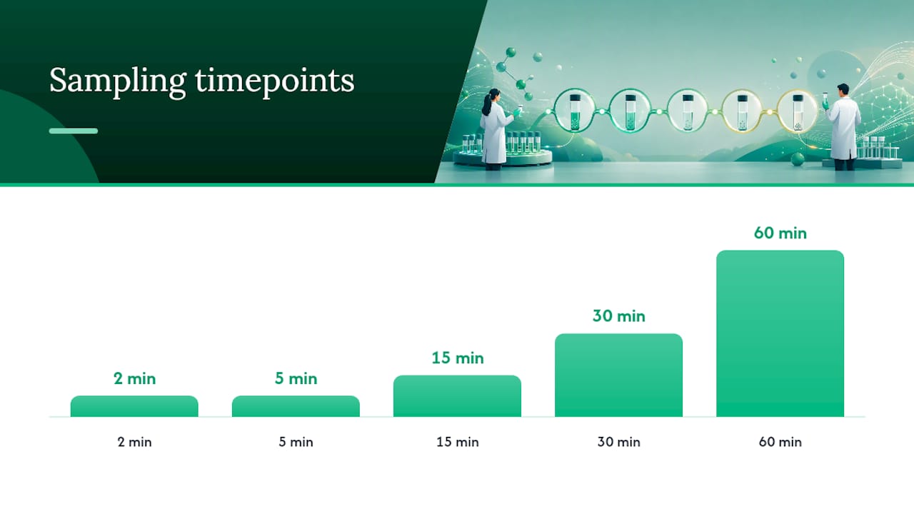
- Sample at multiple early timepoints (0, 2, 5, 15, 30, 60 minutes) since DPP-4 cleavage kinetics are fast enough that a single 24-hour endpoint will miss the informative window entirely.
- Match enzyme concentration to physiologically relevant plasma levels rather than using arbitrarily high enzyme loads that generate artifactual cleavage patterns absent in vivo.
- Confirm truncated product identity by mass shift (loss of His-Ala, roughly 71 Da for the dipeptide minus water) rather than retention time alone, since co-eluting impurities can masquerade as the expected DPP-4 product.
The most common pitfall in enzymatic degradation studies is conflating plasma-based clearance with luminal or tissue-based proteolysis. An oral GLP-1 candidate faces a completely different protease environment, pepsin and chymotrypsin in gastric and intestinal fluid, than an injectable analog circulating in blood. Reviews of marketed GLP-1 receptor agonist metabolism confirm that clearance routes genuinely diverge across analogs: exenatide undergoes renal elimination following hydrolysis, while liraglutide and semaglutide are metabolized through broader proteolytic pathways into constituent amino acids, according to a review of GLP-1 receptor agonist pharmacokinetics. Design your enzyme panel around the delivery route you’re actually targeting, not a generic protease cocktail.
Chemical Degradation: Oxidation, Deamidation, and Hydrolysis Hotspots
Chemical instability operates on a slower but no less damaging timescale than enzymatic cleavage, and it’s the dominant concern once a peptide is sitting in a formulation buffer rather than circulating in plasma. Three residue types carry almost all the risk: tryptophan and methionine for oxidation, asparagine and glutamine for deamidation, and any peptide bond adjacent to aspartate for acid-catalyzed hydrolysis.
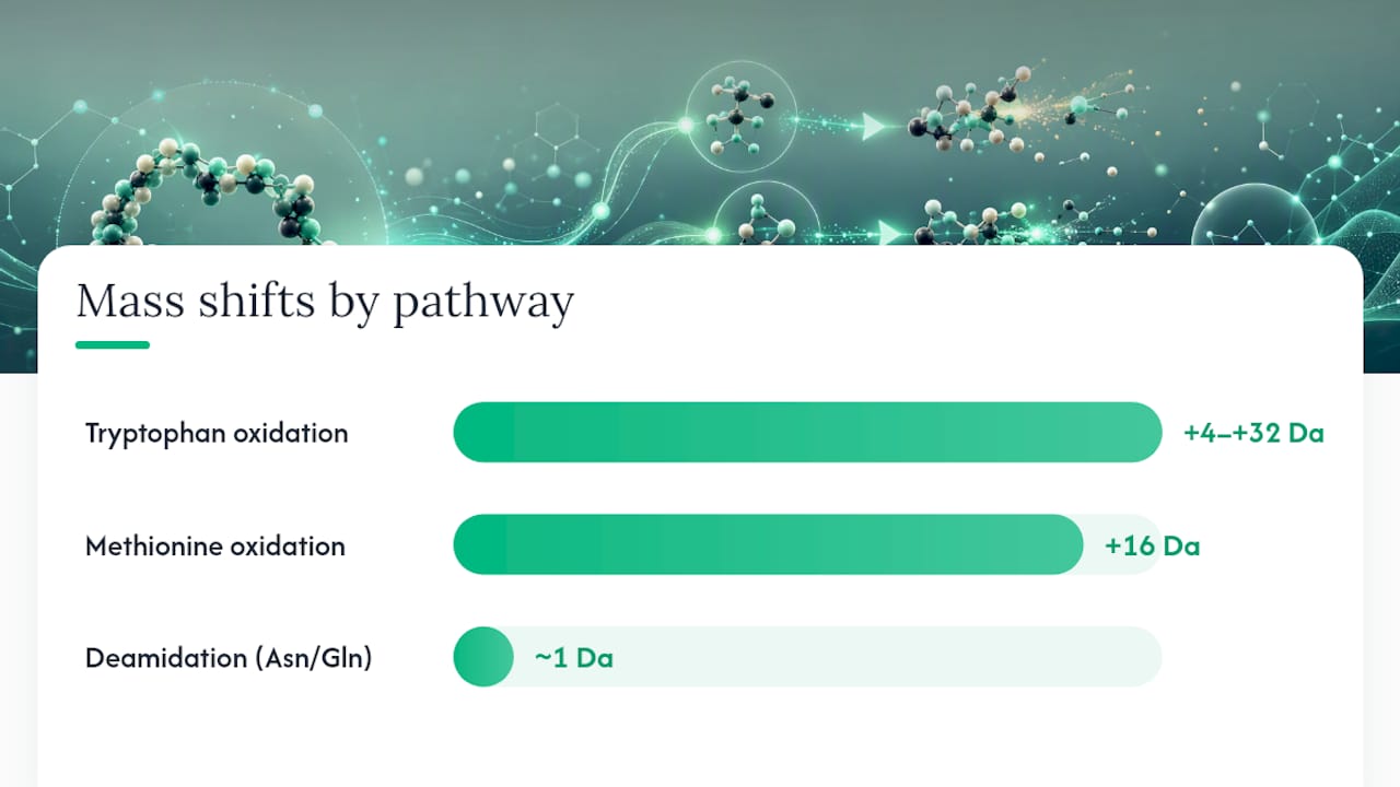
Oxidation of methionine produces a mass shift of +16 Da per oxidized sulfur, detectable by LC-MS as a distinct peak eluting earlier than the parent peptide due to increased polarity. Tryptophan oxidation is messier, generating multiple oxidation products (kynurenine, N-formylkynurenine, hydroxytryptophan) with mass shifts ranging from +4 to +32 Da depending on the extent of oxidation. An application note using LC/Q-TOF MS demonstrated exactly this pattern for GLP-1 agonists stressed with hydrogen peroxide, resolving multiple co-existing oxidized species that would be invisible on a simple UV trace, according to an Agilent method application note.
Deamidation converts asparagine to aspartate or isoaspartate, adding roughly 1 Da (a nearly invisible mass shift that chromatography, not mass spec alone, is needed to detect). The reaction accelerates at alkaline pH and elevated temperature, and it’s often the slow-burn degradation pathway that ruins a six-month stability study nobody flagged at the two-week checkpoint.
Key hotspots and their behavior:
- Trp and Met residues oxidize fastest under light exposure and in the presence of trace metal ions or peroxide contamination in excipients.
- Asn/Gln deamidation rates roughly double for every pH unit increase above neutral, making buffer choice a direct lever on shelf life.
- Asp-Pro and Asp-Gly bonds are unusually acid-labile and can fragment even at moderately low pH during forced-degradation testing.
- Aggregation and oligomerization frequently accompany chemical degradation rather than occurring independently, since partially degraded species often have altered solubility that promotes self-association.
A comprehensive forced-degradation study of liraglutide identified numerous distinct degradation products across acidic, basic, and oxidative stress conditions, with high pH specifically accelerating both aggregation and oxidation in ways that LC-HRMS could resolve and structurally assign, according to research published in the Journal of Peptide Science. That number alone should recalibrate expectations. Researchers who plan for one or two major impurities are going to miss most of what a real stress study produces.
Separately, work on semaglutide’s solution-state stability found that pH, buffer species, molarity, and temperature all independently shift the degradation profile, with certain impurities appearing only in specific buffer systems and disappearing entirely in others, according to a preformulation study using LC-HRMS. That’s a direct warning against generalizing degradation behavior from one buffer system to another, even within the same peptide class. Our semaglutide stability guide walks through specific pH and temperature data if you’re designing a comparable protocol.
Minimizing artifactual chemical degradation during sample handling comes down to a few controllable variables: keep peptide stocks protected from light, add a metal chelator like EDTA where oxidation is a concern, avoid repeated freeze-thaw cycles that promote both oxidation and aggregation, and confirm your buffer’s pH under actual assay temperature rather than at room temperature alone.
Forced-Degradation and LC-MS/MS Workflows for Mapping GLP-1 Breakdown
Mapping how GLP-1 analogs degrade requires a structured stress panel paired with high-resolution mass spectrometry. Guessing at degradation products from a single storage condition produces incomplete, often misleading, stability data.
- Design the stress panel first. Run parallel hydrolytic (0.1N HCl and 0.1N NaOH), oxidative (0.1 to 3% H2O2), and thermal (typically 40 to 80°C) stress arms, sampling at multiple timepoints rather than a single endpoint. Photolytic stress under ICH-standard light exposure is worth adding if the final product will ship in a light-permeable container.
- Separate degradation species by RP-UPLC before mass analysis. A peptide mapping column with a shallow acetonitrile gradient (typically 0.5 to 1% per minute) gives the resolution needed to separate oxidized variants, deamidated species, and truncated fragments that differ by only a few daltons or a single charge state.
- Apply high-resolution MS for accurate mass assignment. Time-of-flight or Orbitrap instruments resolve isobaric degradation products that a lower-resolution quadrupole would report as a single peak.
- Use complementary fragmentation methods for structural confirmation. Collision-induced dissociation (CID) and electron-activated dissociation (EAD) provide different fragmentation patterns; EAD in particular tends to preserve labile modifications that CID can strip away, making the two methods genuinely complementary rather than redundant. Work mapping the oral candidate MEDI7219 used exactly this CID/EAD combination to identify 13 metabolites across 8 distinct cleavage sites, according to research on MEDI7219 metabolite identification.
- Build a parent-to-metabolite map across your time course. Sequential cleavage often shows up as a cascade: an early truncation product becomes the substrate for a second, slower cleavage event. Missing intermediate timepoints can make a two-step degradation pathway look like a single-step one.
- Document findings against ICH Q1A/Q1B and relevant FDA impurity guidance when the goal is regulatory submission rather than exploratory research, since impurity identification thresholds and reporting formats are specified rather than optional.
Pro Tip: Run your forced-degradation panel against a reference standard from the same lot you’re using for the actual assay. Batch-to-batch purity variation, even within a single supplier, can introduce a baseline impurity that gets misattributed to a stress condition instead of pre-existing material.
A comparative study of originator versus analogue liraglutide products found that both showed largely similar impurity profiles under identical stress conditions, reinforcing that comprehensive forced-degradation testing, not a shortened panel, is what actually establishes comparability between a reference product and a research analog, according to a comparative liraglutide degradation study. Skipping stress arms to save time tends to backfire later when an unexpected impurity shows up in long-term storage data with no forced-degradation precedent to explain it.
Sequence Modifications That Improve Proteolytic Stability
Every sequence edit aimed at blocking degradation is a bet against receptor potency. The GLP-1 receptor binding pocket is sensitive enough to N-terminal geometry that even conservative substitutions can shift EC50 by an order of magnitude, so the modifications below need empirical validation, not just structural plausibility, before they’re worth carrying forward.
Substitution strategies with documented stability gains:
- Replacing Ala2 with a D-amino acid or Aib sterically blocks the DPP-4 active site while largely preserving the N-terminal geometry the receptor recognizes.
- Alpha-methylated residues at cleavage-adjacent positions resist chymotrypsin and pepsin specifically, though protection is site-specific rather than universal and requires empirical mapping to confirm, according to a review of peptide backbone modification strategies.
- Lipidation at a non-critical lysine extends half-life primarily through albumin binding rather than direct enzyme resistance, slowing renal clearance and reducing free-peptide exposure to circulating proteases.
- Combining a D-amino acid substitution with lipidation tends to produce additive rather than redundant protection, since the two strategies target different degradation mechanisms.
The MEDI7219 program offers one of the clearer published examples of backbone modification working as intended: alpha-methyl-L-phenylalanine incorporation resisted cleavage by both chymotrypsin and pepsin, and the compound retained roughly 60% of intact peptide after two hours in simulated intestinal fluid, a meaningfully high figure for an oral GLP-1 candidate given how aggressively luminal proteases typically degrade unmodified peptides, according to MEDI7219 metabolite mapping research.
Validation steps that belong in every modification program:
- Confirm GLP-1 receptor activation potency (EC50) in a cell-based cAMP or reporter assay before advancing any modified sequence.
- Run the modified peptide through the same forced-degradation panel as the parent compound to quantify the actual stability gain, not just the theoretical one.
- Use a pharmacokinetic proxy assay (plasma stability incubation, if not a full in vivo study) to confirm that in vitro protection translates to extended systemic exposure.
- Screen for immunogenicity risk when introducing non-natural residues, since D-amino acids and heavily modified backbones can occasionally alter antigen processing in ways a straightforward substitution would not.
There’s a genuine trade-off lurking in all of this: excessive substitution in pursuit of stability risks reducing GLP-1 receptor binding to the point where the compound is more stable but less effective, an outcome that defeats the purpose of the modification in the first place. No sequence edit should advance past initial screening without a head-to-head potency comparison against the unmodified or minimally modified parent peptide.
Formulation Levers That Change Degradation Behavior
Formulation choices act on both enzymatic and chemical degradation simultaneously, often more cheaply than a new sequence modification, which makes formulation screening a reasonable first step before committing to backbone chemistry.
Lipidation’s effect on formulation stability runs in parallel with its effect on plasma half-life. A more heavily lipidated analog binds albumin more tightly, which slows both luminal degradation for oral routes and enzymatic clearance for injectable routes, but it also changes the peptide’s solubility profile in ways that can promote micelle formation or aggregation at certain concentrations.
For oral GLP-1 candidates, permeation enhancers are doing real analytical work, not just improving absorption. Sodium caprate (C10) and SNAC (salcaprozate sodium) both transiently disrupt the intestinal mucosal barrier to improve uptake, and research comparing C10’s effects found it substantially slowed luminal degradation while meaningfully increasing bioavailability in animal models when paired with a stabilized peptide backbone, according to a comparative study on permeation enhancer effects on peptide stability. The enhancer and the sequence modification work together rather than substituting for each other.
Formulation variables worth screening early:
- Buffer species and ionic strength shift the peptide’s oligomeric equilibrium; a small pH change from roughly 6.4 to 8.0 has been shown to meaningfully alter the oligomeric state and degradation profile of liraglutide-type peptides, meaning buffer choice needs to be peptide-specific rather than borrowed from a related molecule.
- Antioxidants (methionine as a sacrificial scavenger, or ascorbic acid at controlled concentrations) reduce oxidative degradation without altering the peptide sequence itself.
- Metal chelators like EDTA limit trace-metal-catalyzed oxidation, a common but underappreciated source of batch-to-batch variability in oxidation rate.
- Surfactants (low concentrations of polysorbate) reduce surface-induced aggregation during agitation or shipping stress, though excess surfactant can introduce its own degradation byproducts over long-term storage.
For prioritization, injectable candidates should weight buffer pH and antioxidant screening heavily since the peptide spends its functional life in a relatively stable, protease-light environment. Oral candidates need the reverse emphasis: permeation enhancer selection and protease resistance testing carry more weight than buffer optimization, since the gut lumen is a far harsher and more variable environment than an injection site.
Preformulation and Storage Practices for Reliable Stability Data
Handling errors, not the underlying chemistry, cause a surprising share of the “degradation” researchers report in bench studies. A rigorous preformulation and storage protocol removes most of that noise before it contaminates real data.
- Select buffer pH relative to the peptide’s isoelectric point (pI), not by default habit. Formulating too close to the pI increases aggregation risk, while formulating far from it in the wrong direction can accelerate hydrolysis at acid-labile bonds.
- Store lyophilized peptide at negative 20°C or below, protected from light and moisture, until reconstitution. Reconstituted solutions degrade markedly faster than lyophilized powder and should be used within the timeframe validated for that specific peptide, not assumed from a related compound. Our lyophilized peptide handling guide covers reconstitution best practices in more depth.
- Aliquot working stocks to avoid repeated freeze-thaw cycles, which mechanically stress the peptide and promote both aggregation and oxidation with each cycle.
- Confirm identity and purity against an authenticated reference standard and its Certificate of Analysis before starting any stability comparison. Batch variation is a real confounder if it goes unchecked.
- Build a documented sample-prep checklist for forced-degradation runs, covering stock concentration, diluent, stress temperature, and sampling schedule, so results are reproducible across operators and instrument runs.
Pro Tip: Run a zero-timepoint control through your entire sample prep workflow, including any dilution and injection steps, before starting a stress study. If you see unexpected degradation products at time zero, the problem is your handling protocol, not the peptide.
Our bench-lab stability rules cover additional detail on minimizing handling-induced artifacts if you’re setting up a new stability protocol from scratch.
A Decision Framework for Balancing Stability and Potency
Choosing between sequence modification and formulation change doesn’t need to be a guess. A structured decision path keeps the trade-offs visible at each step.
- Identify the dominant liability first. Run baseline enzymatic and chemical stress assays to determine whether DPP-4 cleavage, oxidation, or aggregation is driving the majority of observed loss before committing to a fix.
- Try formulation changes before sequence changes when the liability is chemical. Buffer, antioxidant, and excipient adjustments carry no receptor-binding risk and are faster to screen than a new peptide synthesis.
- Reserve sequence modification for enzymatic liabilities that formulation cannot address, since no buffer change will block a protease’s active site.
- Set acceptance criteria before testing, not after. A reasonable starting bar is greater than 90% intact fraction at a defined timepoint, less than 2 to 3 fold EC50 shift versus the parent peptide, and individual impurities below the ICH-recommended identification threshold.
- Flag immunogenicity assessment as mandatory, not optional, whenever a non-natural residue or significant backbone alteration is introduced, since regulatory reviewers treat these modifications as a distinct risk category from conservative substitutions.
- Move to formulation optimization only after the sequence is locked, since re-optimizing formulation around a sequence that later changes wastes both time and material.
The next experiment after initial screening should almost always be a head-to-head forced-degradation comparison between the modified candidate and the parent peptide under identical stress conditions, not a jump straight to long-term real-time stability. That comparison is what tells you whether the modification earned its potency cost.
Why Peptide Purity and Documentation Matter in Degradation Studies
A degradation study is only as trustworthy as the starting material. If a peptide stock carries unknown impurities or an unverified purity claim, every degradation product observed downstream is confounded from the outset, since you can’t distinguish a stress-induced impurity from one that was already present.
A usable Certificate of Analysis should specify the confirmed purity percentage, include the actual HPLC and mass spectrometry traces rather than just a summary number, and identify the specific batch or lot the document corresponds to. Neolabpeptides supplies research-grade GLP-1 analogs and related peptides with third-party HPLC and mass spectrometry verification above 98% purity, shipped lyophilized with a matching COA, intended strictly for laboratory research use and not for human or veterinary application.
A practical incoming-QC checklist before starting any degradation protocol:
- Confirm peptide identity by mass spectrometry against the expected molecular weight.
- Verify purity percentage independently rather than accepting a vendor’s summary claim at face value.
- Check for residual solvents or synthesis byproducts that could interfere with downstream chromatography.
- Compare the COA’s batch number against the physical vial received to rule out documentation mismatches.
Getting this step right before the first stress timepoint saves far more time than it costs.
The Reproducibility Gap in GLP-1 Stability Research
Too much published degradation data comes from studies with slightly different stress conditions, timepoints, and analytical thresholds, which makes cross-study comparison harder than it should be. Standardizing forced-degradation panels, hydrolytic, oxidative, and thermal at minimum, and reporting the full impurity profile rather than just the headline finding, would let the field build on itself instead of re-litigating the same open questions.
The temptation to over-modify a sequence in pursuit of stability is real, and it’s the mistake I’d flag most. A peptide that survives every protease but can’t activate its receptor at a reasonable concentration isn’t a win. Expect the next real progress in this space to come from better analytical sensitivity for low-abundance degradation products and continued work on oral delivery, where the stability problem is hardest and the payoff is largest.
— Stephan
Sourcing Research-Grade GLP-1 Analogs for Stability Work
Degradation studies live or die on the quality of the starting material, and that’s the specific gap Neolabpeptides fills for researchers running these protocols.

The GLP1-S semaglutide analog is available for labs running exactly the kind of forced-degradation and stability comparison work covered above, alongside broader GLP-1 receptor agonist background in our research primer. All products are strictly for laboratory research use, not for human or veterinary application, in line with standard research-chemical regulatory practice. If your protocol requires a custom batch, additional COA detail, or lot-specific documentation for a comparability study, Neolabpeptides can provide it on request. Browse the current catalog and place a research order directly through the site.
Sources
- A comprehensive study on the identification and characterization of major degradation products of synthetic liraglutide using liquid chromatography‐high resolution mass spectrometry
- IDE-mediated GLP-1 degradation as the basis for …
- In‑vitro metabolite identification for MEDI7219, an oral GLP‑1 analog, using LC‑MS/MS with CID and EAD approaches
- Forced Degradation Products of Liraglutide: A Comparative Study of Similarity Between Originator and Analogue Version by Liquid Chromatography–Mass Spectrometry
FAQ
Does GLP-1 lose effectiveness over time?
Yes. Native GLP-1 loses most of its activity within minutes due to DPP-4 cleavage at Ala2, and even stabilized analogs degrade gradually through oxidation, deamidation, and aggregation depending on storage conditions and formulation.
What does GLP-1 analog mean?
A GLP-1 analog is a synthetic peptide engineered to mimic the receptor-activating structure of native glucagon-like peptide-1 while resisting the rapid enzymatic breakdown that limits the natural hormone’s half-life to under two minutes.
Why are some GLP-1 users reporting reduced effects with continued use?
Reported reductions in effect over extended use are more commonly linked to physiological adaptation and dosing factors than to chemical degradation of the drug product itself, though poor storage or handling can independently reduce a formulation’s intact peptide fraction over its shelf life.
Can you give examples of GLP-1 analogs?
Liraglutide, semaglutide, and exenatide are established GLP-1 analogs, each using a distinct stabilization strategy, lipidation, backbone modification, or a naturally DPP-4-resistant exendin-4-derived sequence, to resist degradation.
How do degradation profiles differ between GLP-1 analog drugs?
Degradation routes diverge by analog: exenatide undergoes renal clearance after hydrolysis, while liraglutide and semaglutide are metabolized through broader proteolytic pathways, and each shows a distinct impurity profile under forced-degradation stress testing.
Recommended
This article summarizes published research for informational purposes. Products sold by Neo Lab Peptides are for laboratory research use only and are not intended for human or animal use.