Avoid Missing the 1 nM Peak: GHK-Cu Cell Culture for Labs
September 19, 2026 · STEPHAN ZOHAR

GHK-Cu drives measurable increases in fibroblast proliferation, collagen and elastin synthesis, and glycosaminoglycan production in standard skin cell culture models, typically peaking near 1 nM in a bell-shaped dose response. Gene-expression studies link these outcomes to TGF-β, NF-κB, and antioxidant pathway modulation. For most in vitro protocols, using preformed GHK-Cu with copper-only and vehicle controls produces the most interpretable, reproducible data.
TL;DR:
- GHK-Cu’s optimal effect in cell culture occurs near 1 nM, with higher doses often reducing its benefits due to the bell-shaped dose-response curve.
- It consistently promotes fibroblast proliferation, collagen, elastin, and glycosaminoglycan synthesis, especially in damaged or stressed cells recovering from oxidative or radiation effects.
- GHK-Cu influences gene expression across pathways like TGF-β, NF-κB, and UPS, supporting repair, anti-inflammatory activity, and tissue regeneration, while effects can vary by cell type and conditions.
- Using high-purity, verified GHK-Cu with lot-specific certificates and proper storage enhances reproducibility, especially at low nanomolar concentrations prone to reagent variability.
- Primary fibroblasts are the preferred model for ECM studies, and thoroughly range-finding across multiple concentrations is essential due to the bell-shaped response.
Table of Contents
- What Does GHK-Cu Do to Cells in Culture?
- How Does GHK-Cu Change Gene Expression and Signaling?
- What Concentration of GHK-Cu Works Best in Culture?
- How Should You Design a GHK-Cu Cell Culture Experiment?
- What Results Have Skin Cell Studies Actually Reported?
- Why Does Peptide Sourcing and Purity Matter for GHK-Cu Studies?
- What Are the Biggest Limitations in GHK-Cu Cell Culture Research?
- What I’d Prioritize Before Running a GHK-Cu Study
- Sourcing Research-Grade GHK-Cu With Verified Purity
- Key Primary Sources and Reviews to Consult
- Sources
- FAQ
What Does GHK-Cu Do to Cells in Culture?
The tripeptide glycyl-L-histidyl-L-lysine, bound to copper as GHK-Cu, has been studied in dermal fibroblast and skin models for decades, and the in vitro literature is more consistent than most peptide research. Across multiple independent studies, three effects show up again and again: fibroblasts proliferate faster, extracellular matrix (ECM) output rises, and cells under oxidative or inflammatory stress recover function.
One of the more striking findings comes from irradiated human cervical fibroblast models, where cells exposed to damaging radiation lost their normal growth dynamics. Adding GHK to serum-free medium restored proliferation to levels comparable with non-irradiated controls within one to two days. That is a fast recovery window for a damaged cell population, and it suggests GHK-Cu is acting on core growth-regulatory machinery rather than producing a slow, incidental effect.
The same body of work documents increased synthesis of collagen, elastin, and glycosaminoglycans in fibroblast cultures, alongside stimulated nerve outgrowth, which matters for anyone modeling skin innervation or wound healing in a dish. GHK-Cu also shows anti-inflammatory and antioxidant activity in cultured cells, dampening markers associated with oxidative stress while supporting the structural proteins that give skin its mechanical integrity.
Key observations researchers cite most often in the in vitro literature:
- Increased type I and type III collagen synthesis in human dermal fibroblasts.
- Elevated elastin and glycosaminoglycan production alongside collagen changes.
- Restored proliferative capacity in fibroblasts previously damaged by irradiation or oxidative stress.
- Measurable anti-inflammatory activity, with reduced expression of stress-associated markers.
- Stimulated nerve outgrowth in neuronal and neurite-extension assays relevant to skin innervation.
None of this means GHK-Cu behaves identically across every cell line or passage number. Effect size varies with fibroblast source, culture age, and serum conditions, which is exactly why the mechanistic studies covered next matter for anyone designing a protocol rather than just citing a result.
How Does GHK-Cu Change Gene Expression and Signaling?
The cellular effects above did not emerge from guesswork. They trace back to genomic and pathway-level studies that mapped what GHK actually does inside the cell, and the picture is broader than a single growth-factor bump.
Genomic profiling tied to the Broad Institute’s Connectivity Map found that GHK shifts the expression of a large number of genes involved in tissue repair and inflammation control. This is not a peptide that nudges one receptor. It behaves more like a signaling molecule that resets a broad transcriptional program, which is the mechanistic basis researchers like Loren Pickart have proposed for why aged or stressed fibroblasts shift toward a younger functional phenotype after exposure.
Several signaling axes recur across the mechanistic literature:
- TGF-β pathway activity, linked to increased collagen and ECM gene transcription in fibroblasts.
- Ubiquitin-proteasome system (UPS) activation, which supports clearance of damaged proteins and may partly explain the recovery seen in stressed cell models.
- NF-κB modulation, associated with the anti-inflammatory signal reported across multiple culture studies.
- MAPK pathway involvement, tied to proliferation and survival signaling in fibroblast and keratinocyte models.
These pathways do not operate in isolation, and that is where the phenotype-level story gets interesting. TGF-β activation upregulates collagen and elastin transcription, which shows up downstream as the ECM synthesis increases described in the first section. UPS activation clears misfolded or oxidized proteins, a plausible contributor to the rapid recovery seen in irradiated fibroblast cultures. NF-κB and MAPK modulation together shape whether a stressed cell tips toward inflammation and senescence or toward repair and proliferation, and the in vitro data consistently lean toward the latter under GHK-Cu exposure.
There is a separate and genuinely underappreciated finding worth flagging here: some cancer cell lines show increased apoptotic signaling when exposed to GHK in specific gene-expression contexts, a pattern distinct from the pro-survival effect seen in normal fibroblasts. This is not a contradiction. It reflects the same principle showing up twice: GHK appears to push cells toward whatever transcriptional state is “healthy” for that cell type, which for a stressed but otherwise normal fibroblast means proliferation and repair, and for a dysregulated cancer cell can mean programmed cell death. Researchers working with mixed or transformed cell lines should treat this as a variable to control for, not an inconsistency to explain away.
None of these pathway effects require exotic assay kits. Standard reporter assays, Western blots for phosphorylated MAPK or NF-κB subunits, and qPCR panels for TGF-β target genes are sufficient to capture the signal, provided the concentration range is chosen carefully, which is the subject of the next section.
What Concentration of GHK-Cu Works Best in Culture?
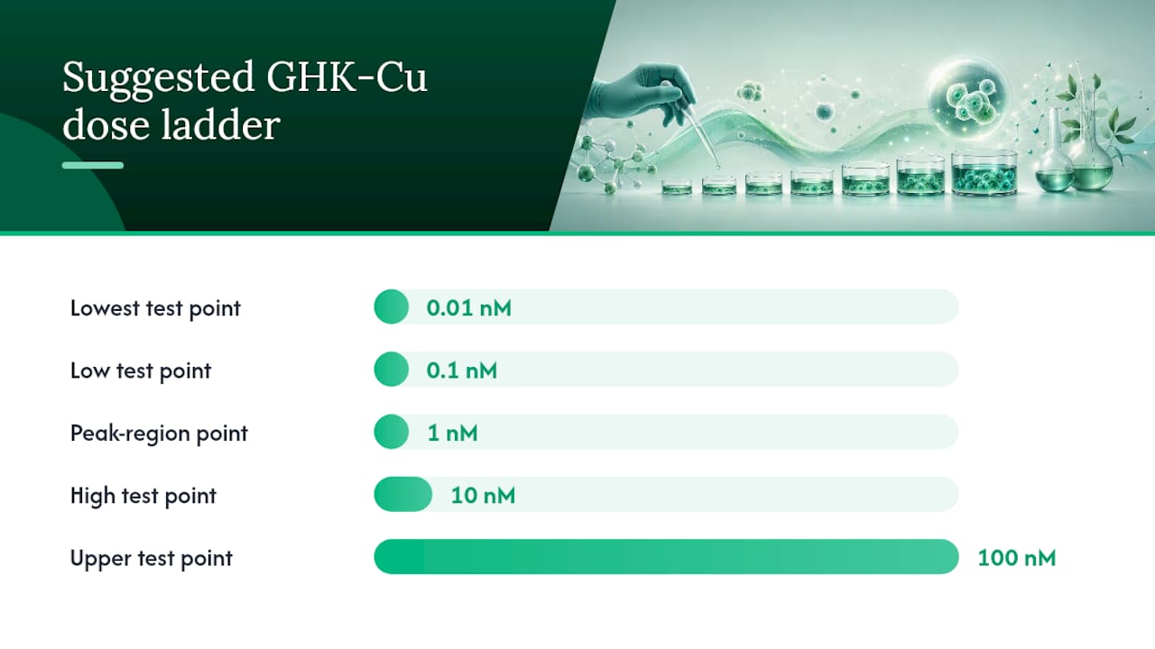
Most in vitro studies test GHK-Cu across a window of roughly 0.01 to 10 nM, with maximal cellular response frequently reported near 1 nM rather than at the highest concentration tested. This is a genuinely counterintuitive feature of the peptide, and it is the single most important design consideration for anyone running a GHK-Cu cell culture experiment for the first time.
The dose-response curve is bell-shaped, not linear. Facial fibroblast studies testing concentrations from 0.01 nM up to 100 nM found that collagen and elastin production, along with favorable MMP and TIMP expression shifts, peaked in the low-nanomolar range and tapered off at higher doses. A researcher who tests only 10 nM and 100 nM, assuming “more is more,” can easily miss the peak entirely and conclude the peptide is weak or inconsistent when the real issue is dose placement.
Pro Tip: Never run a GHK-Cu experiment with just two concentration points. Bell-shaped curves punish sparse sampling; a study with only high and low doses can look like a null result even when the peptide is highly active at the concentration you didn’t test.
Practical steps for range-finding a new GHK-Cu protocol:
- Set a log-scale dose ladder. Use at least five points spanning 0.01, 0.1, 1, 10, and 100 nM to map the full curve on the first pass rather than guessing at a single “working concentration.”
- Run true replicates at each point, not just technical duplicates, since fibroblast response can vary meaningfully between passages and donor lots.
- Include a copper-only control dosed at the same molar copper concentration as your GHK-Cu condition, since copper alone has biological activity that needs to be separated from the peptide’s contribution.
- Include a vehicle-only control using whatever solvent carries your peptide stock, to rule out solvent artifacts in proliferation or ECM assays.
- Repeat the peak-region points (typically 0.1 to 10 nM) in a confirmation run before committing to a single working concentration for downstream experiments.
On the formulation question, GHK-Cu is the endogenous form: the tripeptide has a high natural affinity for copper, and many published protocols use a 1:1 GHK to Cu(II) molar ratio to mirror that physiological complex. That said, several studies achieve clear effects using GHK alone, relying on trace copper already present in standard culture media rather than adding it directly. The practical guidance: if reproducibility with published data matters more than mechanistic purity, use preformed GHK-Cu at a defined ratio. If you’re isolating the peptide’s copper-independent effects specifically, run parallel GHK-only and copper-only arms alongside your GHK-Cu condition. Either way, always report which form you used. “GHK” and “GHK-Cu” are not interchangeable terms in a methods section, even though a lot of published abstracts use them loosely.
How Should You Design a GHK-Cu Cell Culture Experiment?
Protocol design determines whether a GHK-Cu experiment produces clean, citable data or an ambiguous mess that no one else can reproduce. The cell system you choose is the first fork in the road.
Primary human dermal fibroblasts (HDFa) are the workhorse model for ECM and collagen endpoints, and they’re what most of the peak-response data in this field comes from. Keratinocyte lines are the better fit when the research question centers on barrier function or epidermal turnover rather than dermal matrix production. Irradiated or oxidatively stressed fibroblast models are the standard choice for studying GHK-Cu’s protective and recovery effects, since untreated healthy cells simply don’t show much room for improvement. Neuronal or neurite-extension models come into play specifically when the research question involves innervation or nerve outgrowth markers, and they require different media formulations than standard fibroblast work.
Media and passage considerations that affect reproducibility:
- Serum conditions matter enormously for ECM assays. Serum-free or low-serum medium during the treatment window is common in the recovery and proliferation studies, since serum growth factors can mask or compete with the peptide’s own signaling effects.
- Passage number should stay low and consistent across replicates. Primary fibroblasts drift phenotypically after repeated passaging, and a study comparing passage 3 controls to passage 8 treated cells introduces a confound that has nothing to do with GHK-Cu.
- Seeding density needs to leave room for proliferation differences to show up. Overly confluent starting cultures compress the dynamic range of any proliferation assay and can hide a real effect.
- Incubation time depends on the endpoint. Proliferation and viability assays typically resolve within 24 to 48 hours, while collagen, elastin, and MMP/TIMP expression changes are usually assessed at 48 to 96 hours to allow transcriptional and secretory responses time to accumulate.
For assay selection, the field has converged on a fairly standard toolkit. ELISA and Western blot panels for MMP-1, MMP-9, and TIMP-1 are the go-to for matrix remodeling readouts. Hydroxyproline assays or collagen-specific ELISAs quantify total collagen output directly, while Fastin or Sircol-style kits are common for elastin quantification. Proliferation is typically measured with MTT, MTS, or direct cell-count assays, and reactive oxygen species (ROS) levels are captured with fluorescent probes like DCFDA when the research question involves oxidative stress recovery. For neurotrophic outcomes, neurite length and branching under phase-contrast or fluorescence microscopy remain the standard, sometimes paired with qPCR for neurotrophic factor gene expression.
Sample collection timing should be locked to the biological process you’re measuring, not to a single convenient timepoint for the whole experiment. A protocol that collects everything at 48 hours will likely underrepresent early proliferation signals and overrepresent late-stage ECM accumulation, or vice versa. Running a short time-course, even just three collection points across your incubation window, catches this before it becomes a publication problem.
What Results Have Skin Cell Studies Actually Reported?
The MMP and TIMP data give the clearest window into how GHK-Cu affects matrix turnover, because these enzymes control whether collagen and elastin accumulate or get broken down. Studies testing GHK-Cu across low-nanomolar concentrations in human dermal fibroblasts reported increased TIMP-1 expression alongside favorable shifts in MMP activity, a combination that tips the balance toward matrix accumulation rather than degradation. That is the molecular basis for the collagen and elastin increases reported throughout this literature, and it’s a more mechanistically satisfying explanation than simply saying “collagen went up.”
A related dataset extends this into a topical formulation context, where GHK-Cu applied to skin was associated with increased collagen and elastin production alongside measurable reductions in facial wrinkle parameters. It’s worth being precise about what this does and doesn’t tell an in vitro researcher: the wrinkle-reduction endpoint is a clinical, whole-skin outcome, not a cell-culture readout, and it shouldn’t be cited as if it were generated in a dish. The molecular endpoints, MMP/TIMP shifts and collagen/elastin synthesis, are the parts of that study directly comparable to standard cell culture work.
Growth factor production is another consistent thread in the outcomes data. Several fibroblast studies report increased basic fibroblast growth factor (bFGF) output alongside the ECM changes, suggesting GHK-Cu’s effect isn’t limited to direct structural protein synthesis. It also appears to prime fibroblasts to produce more of their own growth signals, which may partly explain why recovery in damaged cell models happens as quickly as it does.
Recurring outcome patterns worth tracking across your own dataset:
- TIMP-1 upregulation correlating with net collagen and elastin accumulation.
- MMP activity shifts consistent with reduced matrix breakdown rather than increased synthesis alone.
- Increased bFGF production accompanying ECM changes in several fibroblast models.
- Nerve outgrowth and neurotrophic marker increases in neuronal co-culture or neurite assays.
- Faster return to baseline proliferation rates in stressed or damaged fibroblast populations compared to untreated controls.
The consistent thread across all of these outcomes is that GHK-Cu’s in vitro effects are matrix-centric and recovery-oriented, rather than acting as a generic growth stimulant. That distinction should shape how you frame your own hypothesis and which endpoints you prioritize.
Why Does Peptide Sourcing and Purity Matter for GHK-Cu Studies?
A dose-response curve is only as trustworthy as the reagent behind it, and GHK-Cu is not a peptide where “close enough” purity is acceptable. A batch with unverified purity, or one contaminated with truncated peptide fragments or free copper in the wrong ratio, will muddy your bell-shaped curve in ways that look like biological variability but are really reagent variability.
Quality benchmark: Research-grade peptides intended for cell culture work are commonly expected to meet greater than 98% purity, verified through HPLC and mass spectrometry, with lot-specific documentation available before the material ever reaches a bench.
Before committing a peptide lot to a full experiment, confirm the following:
- Purity verification via HPLC and mass spectrometry, not just a vendor’s stated percentage without supporting data.
- A Certificate of Analysis (COA) tied to the specific lot number you’re purchasing, not a generic product-page document.
- Molecular weight confirmation matching GHK-Cu’s expected mass, ruling out degraded or incomplete synthesis.
- Clear reconstitution guidance, since improper solvent choice or pH can affect peptide stability before it ever reaches your cells.
Storage matters just as much as initial purity. Lyophilized GHK-Cu should stay frozen and protected from moisture until reconstitution, and once dissolved, it should be aliquoted into single-use volumes rather than subjected to repeated freeze-thaw cycles, which degrade peptide integrity and introduce batch-to-batch inconsistency that can wreck a dose-response study. Neolabpeptides documents purity and identity testing for its GHK-Cu product line with COAs available per lot, and a broader walkthrough of what to check on a COA before you rely on it is covered in this guide to reading a peptide Certificate of Analysis.
What Are the Biggest Limitations in GHK-Cu Cell Culture Research?
Concentration sensitivity is the biggest reproducibility risk in this field. Because the dose-response curve is bell-shaped, small differences in stock preparation, pipetting accuracy at low nanomolar ranges, or even slight variation in a vendor’s reported concentration can shift a result from “clear effect” to “no effect” without any real biological disagreement between labs.
Cell model variability compounds this. Primary fibroblasts from different donors, passage numbers, and even different commercial lots of the same nominal cell line can respond differently to identical treatment conditions. This is a known feature of primary cell work generally, not something specific to GHK-Cu, but it means a single study’s fold-change numbers should be read as a signal, not a fixed constant.
There’s also a meaningful distinction between GHK, GHK-Cu, and lipophilic GHK derivatives that hasn’t been fully mapped in the literature. These forms differ in cellular uptake and stability, and complex culture systems containing skin-derived molecules like urocanic acid can further alter GHK’s effective bioavailability compared to simplified media. Researchers should not assume results from one form or one media system transfer cleanly to another.
Better reporting practices would fix a lot of this. Recording exact lot numbers, publishing COA data alongside results, and specifying the exact assay kit and instrument settings used would let other labs actually replicate a finding instead of guessing at unstated variables.
What I’d Prioritize Before Running a GHK-Cu Study
If I were planning a new GHK-Cu cell culture protocol from scratch, I’d resist the temptation to jump straight to a “known effective” concentration from a single paper. The bell-shaped curve means that number might not transfer cleanly to your cell line, your passage number, or your media formulation. Start with a proper range-finding pass across five log-decades, and treat 1 nM as a hypothesis to confirm, not a given.
Primary human dermal fibroblasts remain the strongest choice for ECM-focused questions, simply because most of the field’s peak-response data comes from that model. If innervation or neurotrophic outcomes matter to your research question, budget separate neuronal co-culture experiments rather than trying to extract nerve-related conclusions from a standard fibroblast assay.
The one habit I’d insist on above everything else: document every lot number and COA alongside your raw data. A peptide’s purity is a variable in your experiment whether you track it or not, and the labs that publish reproducible GHK-Cu work are consistently the ones that treat sourcing documentation as part of the methods section rather than an afterthought in the acknowledgments.
— Stephan
Sourcing Research-Grade GHK-Cu With Verified Purity
Neolabpeptides is built for exactly the reproducibility problem this article keeps circling back to: a dose-response curve is only trustworthy if the reagent behind it is verified, lot by lot, before it reaches your bench.

Every GHK-Cu lot sold through Neolabpeptides ships lyophilized with third-party HPLC and mass spectrometry verification behind the stated purity, and a Certificate of Analysis tied to that specific lot is available before you commit material to a full experiment. That documentation matters most exactly where this article has spent the most time: at low-nanomolar concentrations, where a small purity or concentration discrepancy can shift a bell-shaped curve enough to produce a false negative. The GHK-Cu 100mg product page lists current specifications and COA access, and the broader Anti-Aging & Skin category covers related peptides researchers commonly pair with GHK-Cu work. If you’re building out a range-finding protocol and want to confirm working-stock concentrations before you start, this guide to measuring peptide concentration walks through the methods. For lot-specific COAs, technical specifications, or questions before ordering, reach out through the product page directly.
Key Primary Sources and Reviews to Consult
The GHK-Cu cell culture literature is dense, and reading the full methods sections matters more than trusting any summary, including this one. Start with these:
- The Wiley 2015 review on GHK’s cellular pathways in skin regeneration, for foundational mechanism and fibroblast data.
- The Pickart review on regenerative and protective actions of GHK-Cu, covering gene-data context for nerve outgrowth and ECM effects.
- The genomic modulation paper on GHK and DNA, for Connectivity Map and pathway-level detail.
- The MMP/TIMP and dose-response study, for concentration-specific experimental data.
- Clinicaltrials, to check current translational and clinical trial status directly.
Sources
- GHK Peptide as a Natural Modulator of Multiple Cellular Pathways in Skin Regeneration
- Regenerative and Protective Actions of the GHK-Cu Peptide in the Light of the New Gene Data
- GHK and DNA: Resetting the Human Genome to Health
- Effects of GHK-Cu on MMP and TIMP Expression, Collagen and Elastin Production, and Facial Wrinkle Parameters
- Clinicaltrials
FAQ
Are There Any Human Trials With GHK-Cu?
Registered human trials specifically testing GHK-Cu are limited, and most published evidence for the compound comes from cell culture and topical formulation studies rather than controlled clinical trials. Researchers should search Clinicaltrials directly for the current status of any registered studies before assuming translational data exists.
Is GHK-Cu Risky to Handle in a Lab Setting?
GHK-Cu is a research-use-only compound and carries the standard handling caveats of any laboratory peptide reagent: use appropriate personal protective equipment, avoid ingestion or skin contact with concentrated stock solutions, and follow your institution’s chemical handling protocols. It is not approved for human or veterinary use, and safety data for laboratory personnel should follow standard peptide-handling guidance rather than any therapeutic dosing assumption.
Where Does GHK-Cu Come From?
GHK-Cu is derived from an endogenous human tripeptide naturally found in plasma, saliva, and urine, where it circulates bound to copper. Research-grade GHK-Cu sold for laboratory use, including the version Neolabpeptides supplies, is synthetically manufactured to replicate that native structure rather than extracted from biological sources.
What Are GHK-Cu “Uglies”?
The term sometimes circulates informally to describe transient skin reactions, such as flaking or peeling, reported anecdotally with topical copper peptide use outside of controlled research settings. This has no established basis in the cell culture literature, and researchers should treat it as an internet-forum term rather than a defined scientific phenomenon; any true cytotoxicity or irritation signal should instead be assessed through standard viability assays like MTT or LDH release in your own culture system.
Recommended
This article summarizes published research for informational purposes. Products sold by Neo Lab Peptides are for laboratory research use only and are not intended for human or animal use.